Welcome to the world of dental X-rays, a domain often shrouded in mystery. As a New York cosmetic and general dentist, I’ve seen first-hand the confusion and curiosity surrounding X-rays. So let’s deconstruct this medical marvel together. This blog aims to simplify and decode dental X-rays, offering you a deeper understanding. Prepare to unravel the layers beneath our smiles.
What are Dental X-rays?
Dental X-rays are images of your teeth that your dentist uses to evaluate your oral health. These X-rays are used with low levels of radiation to capture images of the interior of your teeth and gums. This can help your dentist to identify problems, like cavities, tooth decay, and impacted teeth.
Why are Dental X-rays Needed?
Dental X-rays are important for many reasons. They can expose hidden dental structures, malignant or benign masses, bone loss, and cavities. They can also help a dentist prepare for tooth implants, braces, dentures, and other dental procedures. They’re essential in planning and preparing for all types of dental treatment.
Types of Dental X-rays
There are two main types of dental X-rays: intraoral (the X-ray film is inside the mouth) and extraoral (the X-ray film is outside the mouth).
- Intraoral X-rays are the most common type of dental X-ray taken. These X-rays provide a lot of detail and allow your dentist to find cavities, check the health of the tooth root and bone surrounding the tooth, check the status of developing teeth, and monitor the general health of your teeth and jawbone.
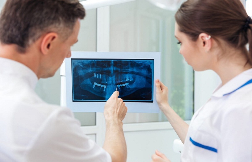
- Extraoral X-rays show teeth, but their main focus is the jaw and skull. These X-rays do not provide the detail found with intraoral X-rays and therefore are not used for detecting cavities or for identifying individual teeth problems. Instead, extraoral X-rays are used to look for impacted teeth, monitor the growth and development of the jaws in relation to the teeth, and identify potential problems between teeth and jaws and the temporomandibular joint (TMJ, the joint that makes the jaw move) or other bones of the face.
Understanding Dental X-rays
Dental X-rays are interpreted and read by a dentist who will look for various conditions such as hidden dental structures, malignant or benign masses, bone loss, and cavities. The interpretation of these X-rays allows the dentist to devise a treatment plan that is suitable for the specific oral health requirements of the patient.
Conclusion
Dental X-rays are a vital part of routine dental care. They offer invaluable insights into the embedded structures of the mouth, which facilitates both diagnosis and remediation. Understanding their use and purpose can help in removing any apprehension, and can lead to improved oral health.